
The Surprising Anatomy of the Turtle
Turtles may look simple from the outside, but their bodies hide some of the most fascinating anatomical features in the animal world. This quick guide helps you understand what makes turtles so unique.
Materials or Tools Needed:
A biology book or reliable online source
Notebook for quick notes
Optional: printed turtle diagram or labeled sketch
Step-by-Step Method:
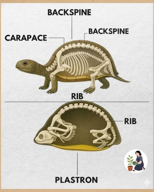
Start by identifying the two main shell parts:
Carapace: the top, hard shell
Plastron: the bottom shell
Note that a turtle’s shell is actually part of its skeleton, fused with the spine and ribcage—meaning turtles can’t leave their shells.
Learn how turtles breathe: unlike mammals, they don’t use a diaphragm. Instead, they use special muscles inside the shell to move air in and out.
Explore their limb anatomy: depending on the species, turtles have flippers (sea turtles) or sturdy legs for walking (land turtles).
Observe how their beak replaces teeth—they use it for biting, scraping, and tearing food.
Organize your notes or diagram to visually map the inside of a turtle for quick understanding.
Additional Tips:
Compare the anatomy of land, freshwater, and sea turtles to see how each adapted to its habitat.
Use color-coded diagrams to memorize shell parts and bone structure.
Watching short documentaries can help you visualize how their internal anatomy works during movement and breathing.